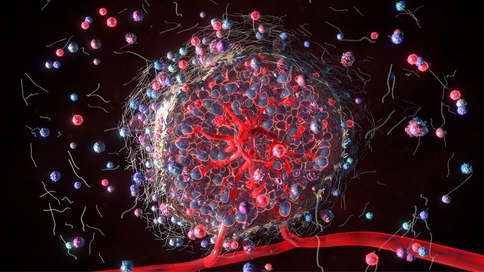
Interrogating the Complexities of the Tumor Microenvironment

Gaining a better understanding of the dynamic and reciprocal interactions between cancer cells and thetumor microenvironment is essential for improving patient diagnosis and treatment.
Cancer cells don’t exist in isolation – instead, they live within a complex ecosystem that also includes immune cells, stromal cells, the extracellular matrix, blood vessels and many other factors. The components of the tumor microenvironment constantly interact and influence each other, which can affect tumor behavior in either positive or negative ways.
“肿瘤细胞可以利用their surrounding microenvironment – by co-opting nutrients and blocking immune surveillance and the host response. But importantly, the tumor microenvironment can also curtail tumor growth and prevent metastasis,” saysJanis Taube, professor of dermatology and pathology and co-director of the Mark Foundation for Advanced Genomics and Imaging at Johns Hopkins University, Baltimore. “It’s a double-edged sword – and so it’s all about getting the balance right and trying to tip it more in favor of the host than the tumor.”
Given the diversity of cell types and molecules that form the tumor microenvironment, it is necessary to apply a systems biology approach to study it.
“Because of the inherent complexity of the tumor microenvironment, changing one thing is going to have ripple effects,” explainsDario A. Vignali临时的椅子和特聘教授Department of Immunology at the University of Pittsburgh and the UPMC Hillman Cancer Center. “You can liken it to a trampoline – if a person stands at one end, there are consequences for anybody else standing on it.”
Dissecting the complexity of the tumor microenvironment may reveal clues for why some cancers respond well to certain treatments while others don’t respond at all or develop resistance over time – and identify potential new vulnerabilities to exploit.
Therapies Targeting Myeloid-Derived Suppressor Cells (MDSCs) in the Tumor Microenvironment

Studying the tumor microenvironment (TME) helps improve our understanding of cancer. Download this whitepaper to learn more about immunotherapy strategies including depletion of MDSC populations and infiltration, inhibition of MDSC recruitment and transport, and anticytokine therapy of MDSCs.
View Whitepaper
Sponsored Content
Targeting the tumor microenvironment
Therapies Targeting Myeloid-Derived Suppressor Cells (MDSCs) in the Tumor Microenvironment

Studying the tumor microenvironment (TME) helps improve our understanding of cancer. Download this whitepaper to learn more about immunotherapy strategies including depletion of MDSC populations and infiltration, inhibition of MDSC recruitment and transport, and anticytokine therapy of MDSCs.
View WhitepaperSponsored Content
In recent years, a new class of immunotherapies that target immune cells within the tumor microenvironment has transformed the treatment of certain types of cancer. Leading this revolution are the so-called immune checkpoint inhibitors that reinvigorate specialized immune cells – called T cells – to recognize, attack and destroy cancer cells.
“These therapies can release the brake that tumors put on the immune system, allowing T cells to respond and help clear the tumor,” explains Taube.
Although some tumors respond well to immune checkpoint blockade, unfortunately, the vast majority are unresponsive. While the reasons behind the high degree of variability between patients are complex, interactions between cancer cells and immune cells within the tumor microenvironment are likely to play an important role.
“There’s a variable degree of immune infiltration in tumors, which has given rise to the analogy of an ‘immune desert’ or an ‘immune jungle’,” describes Vignali. “There’s good data to suggest that tumors that are highly immune infiltrated are more sensitive to the effects of these immunotherapies.”
Researchers are striving to define the features of the tumor microenvironment that influence how well a person’s cancer responds to immune checkpoint inhibitors – to identify new biomarkers for personalizing treatment and find ways to boost the effectiveness of these drugs.
“We need to understand the effect of these treatments on the entire tumor microenvironment, rather than only focusing on the specific type of cell they’re targeting,” says Vignali. “A killer T cell isn’t there in isolation – it’s interacting with lots of other cells, which will have knock-on effects on how the tumor responds to checkpoint inhibition.”
Precise Spatial Multiplexing of Immune Cell Diversity in Clinical Tumor Samples

Download this poster to discover a platform that can generate highly-multiplexed, spatially-resolved protein expression data from a clinical sample, quantify cell populations expressing very high or low levels of a single marker and use commercial antibodies from any vendor to spatially resolve protein targetsin situ.
View Poster
Sponsored Content
Precise Spatial Multiplexing of Immune Cell Diversity in Clinical Tumor Samples

Download this poster to discover a platform that can generate highly-multiplexed, spatially-resolved protein expression data from a clinical sample, quantify cell populations expressing very high or low levels of a single marker and use commercial antibodies from any vendor to spatially resolve protein targetsin situ.
View PosterSponsored Content
Single-cell technologies
Until recently, researchers were limited to profiling whole tumor samples using ”bulk sequencing” approaches. But the advent of novel single-cell technologies is enabling them to profile immune cell populations within the tumor microenvironment in more granular detail.
“We can get an enormous amount of information from just one cell – it’s been game-changing,” enthuses Vignali.
Single-cell transcriptomics enables the analysis of the abundance and sequences of RNA molecules, while epigenomics is the genome-wide mapping of DNA methylation, histone protein modification, chromatin accessibility and chromosome conformation. Another recent innovation is cellular indexing of transcriptomes and epitopes by sequencing (CITE-seq), which enables researchers to simultaneously capture RNA expression and cell surface expression of specific proteins on the same cell. Sequencing immune cell receptors – such as the T-cell receptor (TCR) and the B-cell receptor (BCR) – can provide a further dimension of information for characterizing their specificity, function and phenotype.
“For instance, we think of killer T cells as being one group – but in reality, when you examine them at the single cell level, you will find that there are multiple, distinct subsets within that population,” says Vignali.
Applying multi-omics single-cell technologies is allowing researchers to ask questions about the tumor microenvironment that were previously out of reach.
“We’ve always been limited by the amount of tissue you can get from a patient – but it’s now possible to get a lot of information even from a small tumor biopsy,” says Vignali. “For instance, we have an ongoing clinical trial where we’re collecting core tumor biopsies from patients before and after treatment. It’s not an overly invasive procedure and it means we can look at the baseline situation and then what changed after immunotherapy.”
High Resolution Mapping of the Breast Cancer Tumor Microenvironment

Download this app note to learn how a single cell gene expression workflow was used to analyze large, serial formalin-fixed, paraffin-embedded human breast cancer sections, identify and spatially resolve 17 different cell types and their gene expression profiles, and spatially register protein, histological and RNA data together into a single image.
View App Note
Sponsored Content
High Resolution Mapping of the Breast Cancer Tumor Microenvironment

Download this app note to learn how a single cell gene expression workflow was used to analyze large, serial formalin-fixed, paraffin-embedded human breast cancer sections, identify and spatially resolve 17 different cell types and their gene expression profiles, and spatially register protein, histological and RNA data together into a single image.
View App NoteSponsored Content
Mapping the tumor microenvironment
However, while single-cell technologies can shed light on the molecular characteristics of the cells that make up a tumor, they don’t provide any information about how cells are organized spatially within their microenvironment.
Multiplex immunofluorescence (mIF) techniques are becoming an increasingly important tool for analyzing multiple cell types and their geographic relationships within the tumor microenvironment. The approach enables the simultaneous antibody-based detection of multiple markers on a single tissue section, but the analysis and visualization of mIF data can be complex and time-consuming. But lessons can be learned from the techniques already used by astronomers to map the universe.
“Our big realization is there are many similarities between describing the celestial sphere and annotating the tumor microenvironment,” saysAlexander Szalay, Bloomberg distinguished professor of physics and astronomy at Johns Hopkins University. “The bottom line is that everything is about characterizing spatial relationships – so what are the objects in proximity that are interacting with each other, and how can we represent this visually?”
A unique collaboration forged between Taube and Szalay led to the development of theAstroPath platform, which enables the comprehensive analysis of multispectral imaging datasets in tumor tissue sections at single-cell resolution.
“We use immunofluorescent tags on antibodies to label multiple markers on a section – and we can then identify every single cell,” explains Taube. “Using this approach, we can reconstruct multispectral images from across a whole slide, giving us around a hundred times more data about the tumor microenvironment than was available previously.”
The pair are using this formidable technology to advance the understanding of the interplay between cancer cells and the immune system and to identify predictive biomarkers of the tumor response to immunotherapies. The development of the platform as an open resource for visualizing and analyzing spatially resolved mIF datasets is already underway.
“TheSloan Digital Sky Survey, which Alex helped to architect and curate, has had over three billion hits to its website to date,” says Taube. “We hope to make the tumor microenvironment equivalent of that resource, which will allow people all over the world to peruse and query these data.”
Understanding Tumor Microenvironment Signaling Pathways

肿瘤微环境(时差)是由the tumor, extracellular matrix and various non-transformed cells including fibroblasts, immune infiltrates and vascular vessels. Download this eBook to learn more about receptor tyrosine kinase signaling, cytokine receptors and ancient signaling.
View eBook
Sponsored Content
Understanding Tumor Microenvironment Signaling Pathways

肿瘤微环境(时差)是由the tumor, extracellular matrix and various non-transformed cells including fibroblasts, immune infiltrates and vascular vessels. Download this eBook to learn more about receptor tyrosine kinase signaling, cytokine receptors and ancient signaling.
View eBookSponsored Content
Exponential progress
Understanding how the dynamic interactions of cancer cells with their surrounding microenvironment can influence tumor behavior, prognosis and response to treatment could ultimately help transform patient care.Thanks to huge advances in technology, researchers are now equipped with the tools to study the tumor microenvironment in more detail than ever before.
“These interrogative technologies have opened the door to gain substantially more information at an exponential rate over the next decade or so,” says Vignali. “The capabilities are enormous – and it’s almost a cliché, but the sky really is the limit!”
However, this is presenting new challenges with integrating and interpreting the multiple enormous datasets generated – leading some researchers to seek solutions from unexpected sources.
“I spent decades in astronomy working on these special tools – and I never thought this would have a practical application,” says Szalay. “And now it’s clear that one day, this work may actually save lives – and that’s incredibly rewarding.”


